Work
Research projects in AI for medical imaging, alongside software I build in my own time. Pick a project to read the full story.
AMARA — AI for lung nodule malignancy estimation
- Python
- PyTorch
- NumPy
- pandas
- scikit-learn
- Git
Deep learning has transformed medical image analysis. Radboudumc developed an algorithm for pulmonary nodule malignancy prediction that matches the performance of thoracic radiologists on low-dose chest CT.
To bring this algorithm into routine clinical practice, we are enhancing it with additional CT data and validating it against data from European lung cancer screening trials and five Dutch institutes. The ultimate goal is an AI tool that speeds up the diagnosis of malignant nodules and reduces unnecessary examinations of benign ones. A full description of my PhD project is available at DIAG Nijmegen.
The LUNA25 challenge
The LUNA25 challenge is a benchmarking initiative that evaluates AI algorithms and radiologists on lung cancer screening in CT. It provides a standardized dataset and evaluation framework to assess the accuracy, sensitivity, and specificity of AI models in detecting lung nodules and predicting malignancy risk. By comparing algorithms directly with human readers, the challenge advances the development of reliable AI tools for lung cancer screening.
Uncertainty estimation
Safe clinical implementation of a deep learning algorithm requires the ability to gauge and communicate uncertainty. Uncertainty estimation identifies situations where the algorithm has doubts about its prediction, so those cases can be deferred to clinical experts instead of being decided automatically. This proactive integration minimizes algorithm mistakes and raises the safety profile of a clinically adopted model.
Out-of-distribution detection
Deep learning models can fail silently when applied to data that differs from their training distribution — a phenomenon known as dataset shift, caused by changes in patient demographics, disease incidence, or image acquisition and reconstruction. Incorporating an out-of-distribution detection component helps identify these shifts and supports safe, reliable clinical deployment.
Kilometer tracker
- Python
- Django
- HTML5
- CSS
- Git
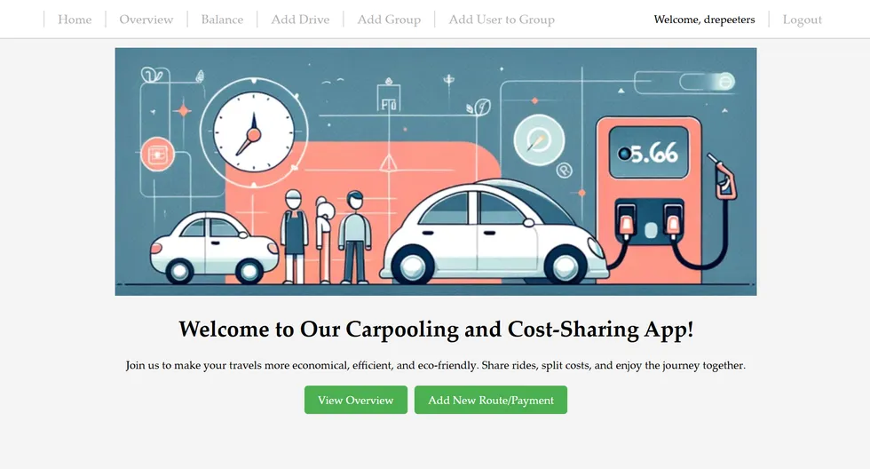
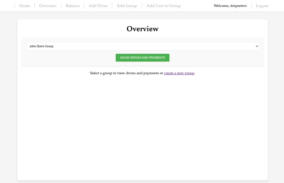
Sharing a car with someone close, like a partner or colleague, made me realize how important it is to track the kilometers driven by each person and distribute fuel expenses fairly. That inspired me to build a practical, user-friendly kilometer tracker. The application lets users register their trips, monitor kilometers driven, and keep track of fuel payments, ensuring transparency and fairness.
Built as a real-world solution, the project combines Python and Django on the backend with plain HTML and CSS on the frontend, and is hosted online for seamless access.
Outcome
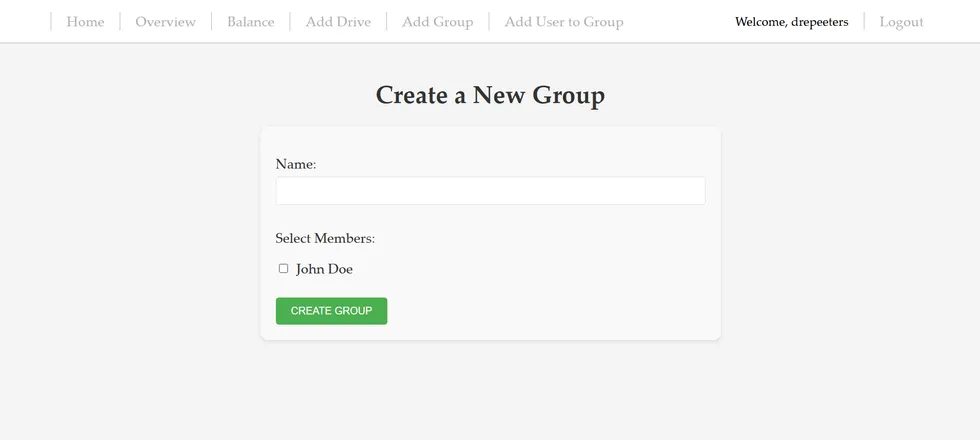
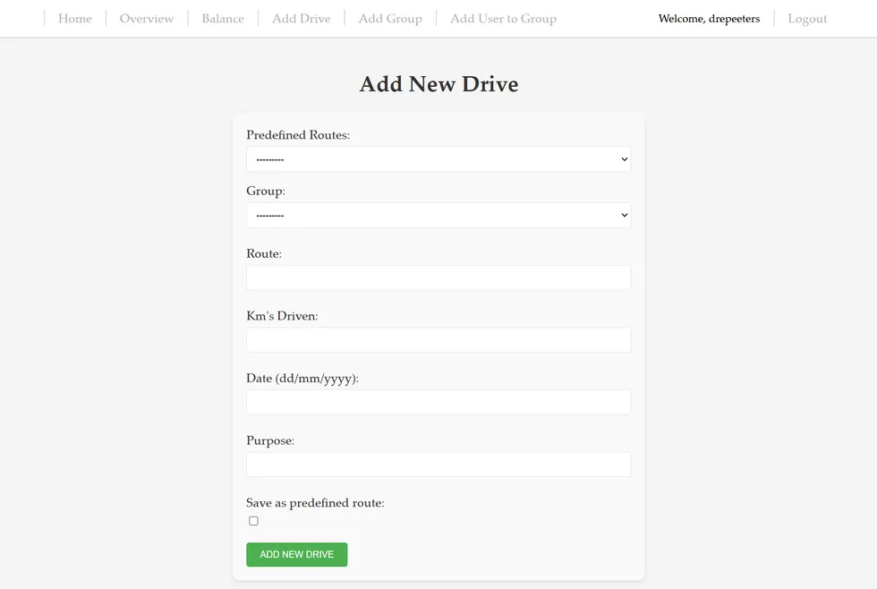
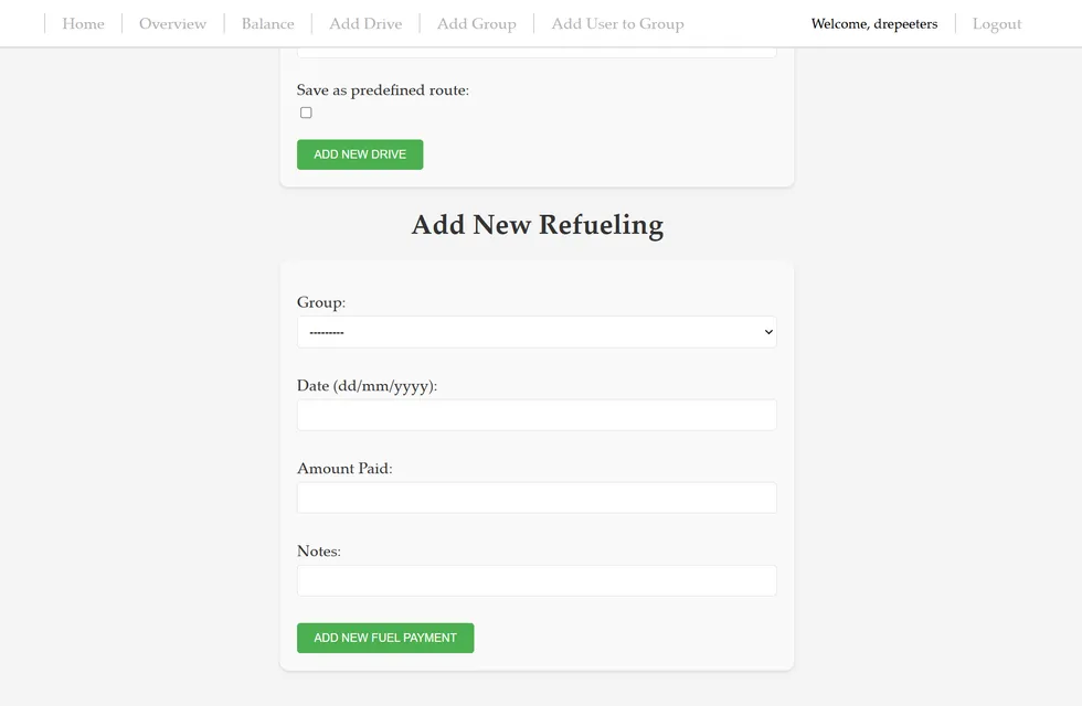
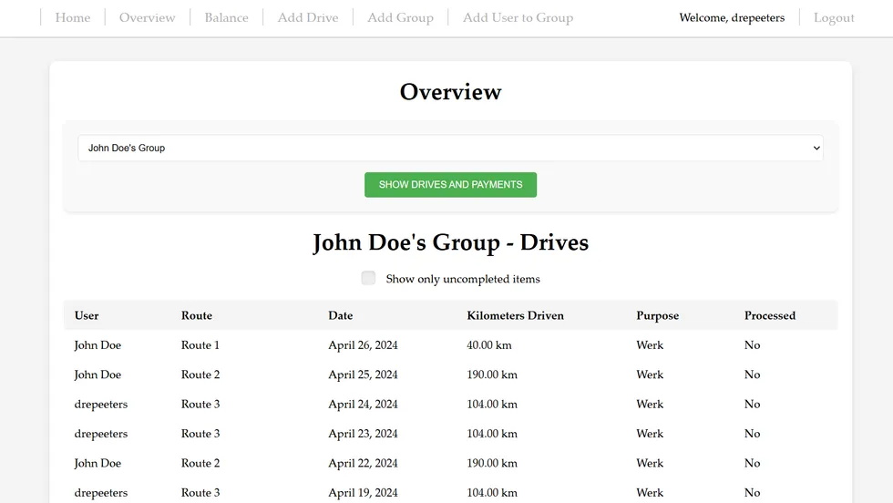
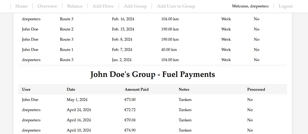
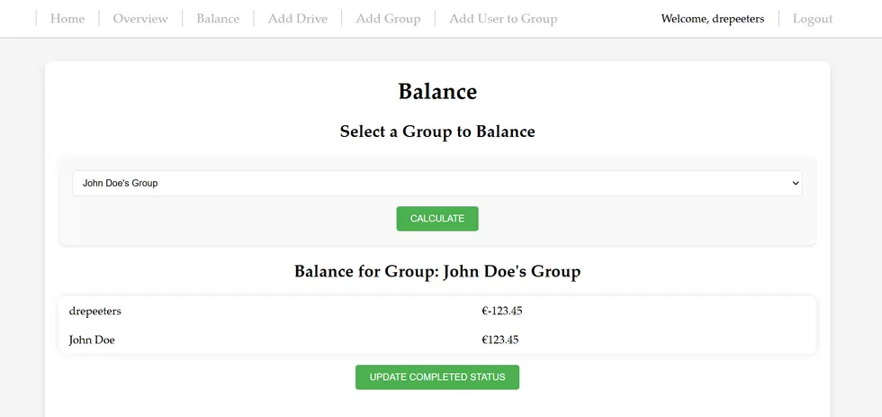
A fully functional online application that simplifies trip and expense tracking for car-sharing users. Its features include:
- User authentication
- Creating user groups
- Registering trips and payments per group
- An overview of all trips and payments
- An option to balance the group
Automated X-ray quality control
- Python
- PyTorch
- NumPy
- Git
The LRCB is the Dutch national expert center that performs quality control of the medical devices used in the national screening programs for breast cancer and tuberculosis. The current quality control procedure for phantom images from mammographic devices is time-consuming and observer-dependent: physicists manually inspect phantom images for visible artefacts.
Experiments
We used several pretrained deep learning models to classify phantom images, both as a binary task (visible versus no visible artefact) and as a multi-class task distinguishing eight artefact classes from artefact-free images.
Outcome
The trained models classified artefacts accurately in both the binary and the multi-class setting. These results demonstrate the potential of deep learning in the quality control of X-ray devices.
Microcalcification malignancy risk estimation
- Python
- scikit-learn
- pandas
- NumPy
- Git
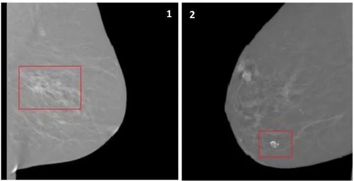
In the diagnosis of microcalcifications linked to breast cancer, most patients undergo a biopsy because radiologists find it challenging to reliably discern benign from malignant lesions. Most of these biopsies turn out benign, causing unnecessary stress for patients and increased diagnostic costs.
Experiments
We extracted radiomics features from microcalcifications on low-energy contrast-enhanced spectral mammography images and developed multiple machine learning models, trained both with and without clinical parameters.
Outcome
The models showed excellent discriminative performance in classifying benign and malignant microcalcifications, demonstrating their potential to improve clinical care and reduce unnecessary biopsies.
Balance — recipes and meal planning
- React
- JavaScript
- Python
- Git
Keeping track of recipes, planning meals, and doing grocery shopping can be time-consuming and overwhelming. To address this, I am developing a mobile recipe management and meal planning application using JavaScript, React, and Python.
Outcome (in progress)
A recipe management and meal planning application with an AI-powered chat bot that helps users track their recipes, plan meals, and generate shopping lists based on selected recipes.